
3.68
new VTE cases per 100 resident-years
Reported across 181 nursing homes in 19 U.S. states.
Wireless Ultrasound for Suspected Deep Vein Thrombosis
DVTScanner™ brings portable ultrasound capability, implementation support, and a clinician-directed workflow closer to patients. Choose the path for Post-Acute & Long-Term Care or Primary Care.
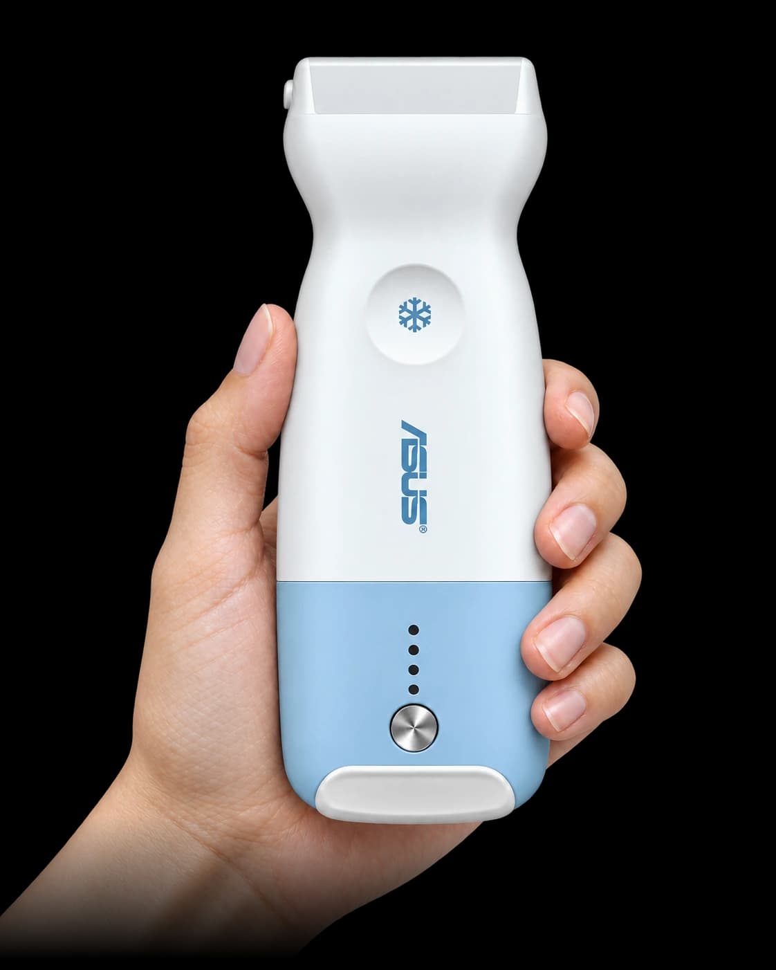
DVTScanner™ is a Focused POCUS Solution from

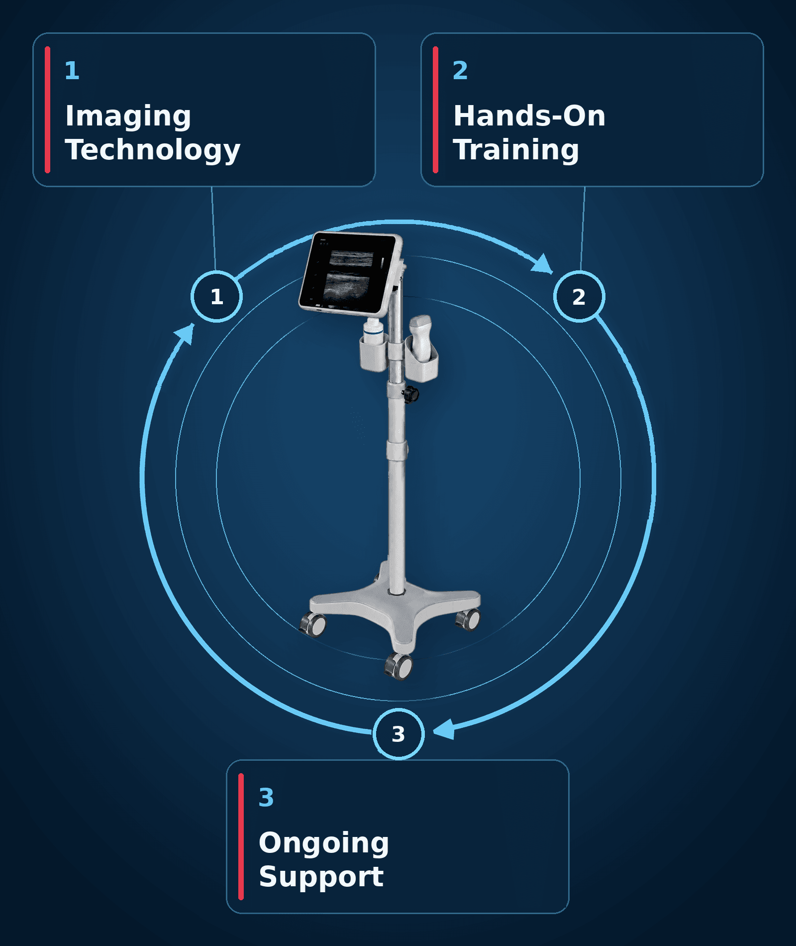
The Complete Solution
We help you implement a DVT program with a complete solution built around three essential elements.
ASUS LU800 wireless linear ultrasound with a fully configured mobile cart.
ARDMS- or CCI-registered sonographers (RVT, RDMS, or RVS) train your clinicians on-site to scan, capture images, and follow the DVT workflow.
Your team receives refresher training, continuing education, and ongoing help.
How It Works
The DVTScanner workflow follows the same three steps in a nursing facility or a primary care office. Who performs each step is set by your organization’s policies and each team member’s scope of practice; we map it with you during the pilot.
A resident or patient has a swollen or painful leg, or another finding that raises concern for DVT. The provider orders a bedside scan the way they would order any point-of-care test.
A trained member of your team brings the cart to the bedside or exam room and captures a proximal venous compression study using the protocol taught in training. The study takes minutes.
The provider reviews the study and decides the next step: begin treatment, order a confirmatory venous duplex when clinically indicated, or manage a negative scan by the patient’s pretest probability under your protocol. For a higher-risk patient that can mean a repeat scan in about a week.
The role map for your setting is confirmed in your pilot discussion.
The Population At Risk
3.68
Reported across 181 nursing homes in 19 U.S. states.
Within 7 days
In a Minnesota population-based study of 3,465 nursing-home residents.
About ½
A critical transition period for many post-acute residents.
Rates are population findings and do not predict an individual resident’s risk. Clinical evaluation and appropriate testing remain essential.
The Delay Problem
When symptoms arise, the path to traditional imaging requires coordination, delay, or transport.
Every hour between suspected DVT and a confirmed diagnosis can delay treatment decisions. For long-term-care residents, the traditional path often means scheduling a mobile vendor or arranging transport, then waiting for the images to be interpreted and reported. Bedside scanning brings imaging to the resident and helps shorten that path.
The CMS standard
>24 h
For this measure, CMS1173 (DOVE) counts a VTE diagnosis made more than 24 hours after the primary-care visit where symptoms were first documented as delayed.
How often that line is missed
79-82%after 24 h
70-71%after 72 h
Across 3,525 patients at two U.S. health systems, delayed diagnosis was common. At the larger system, 30-day mortality was 8.33% with delayed diagnosis versus 2.52% with timely diagnosis, a risk ratio of 3.31.
A documented care delay
Friday → Monday
A Minnesota Department of Health investigation of an assisted-living facility documented a family member being told on a Friday that an ultrasound to check a resident’s swollen leg for a blood clot would wait until Monday.
The CMS and JAMA findings describe primary-care populations. The Minnesota investigation documents one assisted-living resident’s experience. Its finding concerned missed medication, and it does not show that the wait for the ultrasound caused harm.
Primary Care
Primary care has evidence for both sides of the problem. CMS1173 defines a VTE diagnosis made more than 24 hours after a primary-care visit where symptoms were documented as delayed, and at two U.S. health systems most qualifying cases met that definition.
Primary care also has direct evidence that trained physicians can perform proximal compression ultrasound. In Italy’s multicenter PRACTICUS study, general practitioners reached 95.8% diagnostic accuracy, with excellent agreement with blinded vascular-ultrasound experts (κ = 0.86).
2023 · DOVE beta test
72.6%Site 1
77.1%Site 2
At two U.S. health systems, nearly three in four qualifying VTE cases met the DOVE definition of delayed diagnosis. Across the 50 largest primary-care sites at Site 1, rates ranged from 46.15% to 100%.
2025 · DOVE follow-up
79-82%after 24 h
70-71%after 72 h
Across 3,525 patients at two U.S. health systems, delayed diagnosis was the norm. At the larger system, 30-day mortality was 8.33% with delayed diagnosis versus 2.52% with timely diagnosis, a risk ratio of 3.31.
The next measurement phase
$2 million
Grant R01 HS030221 is extending DOVE across urban and rural primary and urgent care, identifying causes of delayed and missed VTE diagnoses, and measuring their costs.
PRACTICUS · Italy
95.8%
After two months of formal training (12 hours didactic, 30 hands-on, 8 image review), 18 general practitioners performed proximal compression ultrasound in 1,107 consecutive outpatients: 90.0% sensitivity, 97.1% specificity, and κ = 0.86 versus blinded vascular experts.
PRACTICUS evaluated trained physicians performing bilateral proximal compression ultrasound. It does not establish autonomous diagnosis or replace comprehensive venous duplex when clinically indicated.
Sources
Start a Pilot
Talk with CorVascular about your patient population, current diagnostic pathway, staffing model, and pilot objectives.
Program pricing and billing guidance by setting are covered in your pilot discussion.
DVTScanner™ is a trademark of CorVascular. ASUS® is a registered trademark of ASUSTeK Computer Inc. Intended for healthcare professionals. Use of ultrasound for DVT assessment is subject to each organization’s clinical policies and the operator’s scope of practice.
CorVascular is the exclusive U.S. importer and vascular market distributor for ASUS's portfolio of FDA-cleared ultrasound devices.